ConRad MRI Safety Testing Texas Childrens
1.Background
2.Testing Setup
3.Protocol
4.Results
5.Conclusions
6.Limitations
1.Background
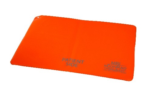
The patients under HIFU procedure loss their temperature very quick due to convection and conduction and radiation. We have purchased a special thermal blanket to keep patients warm. The thermal blanket, bought from Veterinary Warming Solutions LLC, is labeled as ConRad (MRI-Safe) Technical Information. There are two component layers inside: 1) a heat insulative layer made of Rayon 2) a heat trapping layer made of polyurethane. Therefore, there are no ferromagnetic and conductive material to cause hazards in the MR environment. However, since the company haven’t test it on a scanner for human, we are going to perform a test focusing on artifacts and possible heating related safety on our clinic 3T Siemens Skyra scanner.
2.Testing Setup
Phantom: A 5 liters phantom was wrapped inside the ConRad thermal blanket and put in the center of a 20-channel head coil. The temperature probe was firmly attached to the surface of the phantom using tapes. The temperature changes is displayed on the monitor. The setup is shown in the following figure:


Volunteer: Second part was the tests on a volunteer with 178 cm height and 82 kilogram weight. The blanket was set on top of the volunteer’s chest since its size is not large enough to wrap around the participant body. This would be a significant difference between our test and what is expected in the reality of HIFU procedure on pediatric patients. The temperature probe was put under armpit for recording the temperature change.
3.Protocol
Phantom: Two sequences was used in the testing: 1. DWI epi sequence for testing of artifacts. 2. T2 space for testing heat deposition.
| Institution Name | Texas Childrens Main | Texas Childrens Main | Texas Childrens Main |
| Manufacturer's Model Name | Skyra | Skyra | Skyra |
| Software Versions(s) | syngo MR E11 | syngo MR E11 | syngo MR E11 |
| Series Description | localizer_quiet | DWI ep2d_diff_orth_p2 | AX T2 SPACE |
| Series Number | 1 | 2 | 5 |
| Number of Averages | 2 | 4 | 12 |
| Repetition Time | 10 | 5900 | 1400 |
| Echo Time | 3.69 | 102 | 155 |
| Flip Angle | 20 | 90 | 135 |
| Slice Thickness | 7 | 4 | 0.600000024 |
| Rows | 512 | 192 | 384 |
| Columns | 512 | 192 | 384 |
| Number of Phase Encoding Steps | 233 | 143 | 304 |
| Echo Train Length | 1 | 71 | 69 |
| Percent Sampling | 91 | 100 | 100 |
| Percent Phase Field of View | 100 | 100 | 100 |
| Pixel Spacing | 0.48828*0.48828 | 1.1458*1.1458 | 0.57292*0.57292 |
| MR Acquisition Type | 2D | 2D | 3D |
| Imaging Frequency | 123.262462 | 123.262514 | 123.262507 |
| Pixel Bandwidth | 355 | 1040 | 290 |
| SAR | 0.016362395 | 0.135543143 | 0.507811481 |
| duration (minutes) | 2 | 1.2 | 44.4 |
Volunteer: The testing sequences includes high SAR T2 TSE, EPI DWI, EPI fMRI and fieldmap, aiming to find out any discomfort and image artifacts caused by the blanket. Totally it takes around 10 minutes.
| Institution Name | Texas Childrens Main | Texas Childrens Main | Texas Childrens Main | Texas Childrens Main | Texas Childrens Main |
| Manufacturer's Model Name | MAGNETOM Vida | MAGNETOM Vida | MAGNETOM Vida | MAGNETOM Vida | MAGNETOM Vida |
| Software Versions(s) | syngo MR XA20 | syngo MR XA20 | syngo MR XA20 | syngo MR XA20 | syngo MR XA20 |
| Series Description | ead_scout_64ch-head-coil_MPR | AX DWI_TRACEW | AX T2 | gre_field_mapping | measurement functiona |
| Series Number | 3 | 5 | 7 | 8 | 10 |
| Number of Averages | 1 | 2 | 2 | 1 | 1 |
| Repetition Time | 3.15 | 7000 | 6430 | 400 | 3000 |
| Echo Time | 1.37 | 114 | 95 | 4.92 | 30 |
| Flip Angle | 8 | 90 | 150 | 60 | 90 |
| Slice Thickness | 1.6 | 4 | 4 | 3 | 3 |
| Rows | 162 | 296 | 512 | 94 | 94 |
| Columns | 162 | 292 | 512 | 94 | 94 |
| Number of Phase Encoding S | 160 | 148 | 358 | 94 | 94 |
| Echo Train Lengthssss | 1 | 73 | 13 | 0 | 47 |
| Percent Sampling | 100 | 100 | 70 | 100 | 100 |
| Percent Phase Field of View | 100 | 101.37 | 100 | 100 | 100 |
| Pixel Spacing | 1.6*1.6 | 0.85616*0.85616 | 0.48828*0.48828 | 2.6596*2.6596 | 2.6596*2.6596 |
| MR Acquisition Type | 3D | 2D | 2D | 2D | 2D |
| Imaging Frequency | 123.243655 | 123.243679 | 123.243655 | 123.243674 | 123.243681 |
| Pixel Bandwidth | 539 | 1631 | 195 | 598 | 1773 |
| gain | 391.561 | 391.561 | 391.561 | 391.561 | 391.561 |
| SAR | 0.028502319 | 0.173783505 | 0.541391252 | 0.157953401 | 0.138823148 |
| duration (min:sec) | 1:30 | 2:30 | 5:15 | 1:42 | 0:45 |
4.Results
Phantom: There was no extra artifact found on images of the DWI EPI sequence (see figure left)
The temperature gradually increased along with time during the 45 minutes continue scan. 
The temperature increased at a rate of 0.04 (oC) per minute. The maximum increase of temperature was 1.7 (oC) within 45 minutes continue scan. This was normal energy deposition by a high SAR sequence. The blanket didn’t raise any concern of MR safety.

Volunteer: The temperature increased within one degree Celsius from 34.9 oC to 35.5 oC. No notable extra artifacts were found on fieldmap, DWI and fMRI images:

According to the Volunteer, there was no discomfort during the MR session.
6.Limitations
Our volunteer test was performed on adult which is very different from an infant in our HIFU study in terms of SAR and artifacts.
5.Conclusions
Since no ferromagnetic and no metal components was contented in the blanket according to the description from the manufacture in combination of our tests, it is safe to conclude that the ConRad thermal blanket should be safe in our HIFU 3T MR system.
-
ConRad Thermal Blankets
Regular price From $79.95Regular priceUnit price per -
ConRad Thermal Blankets Complete Set
Regular price $242.95Regular priceUnit price per -
MRI Safe - ConRad Thermal Blankets
Regular price From $114.95Regular priceUnit price per -
MRI Safe - ConRad Thermal Blankets Complete Set
Regular price $419.85Regular priceUnit price per